PRP Physio logo

Hip Pain Physiotherapy Brisbane

Hip Injury Rehabilitation

Hip pain is frustrating, trust me, I experienced it for about 2 weeks earlier this year and it sucked. Putting on pants was a struggle because I just couldn’t stand on that leg without either falling over or being in excruciating pain. I only experienced it for about 2 weeks, so I’d hate to have lived with that for months or even years, as some of my clients have.

Hip pain presentations can change a lot throughout the lifetime, from the gluteal tendinopathy that I experienced, to trochanteric bursitis, through hip osteoarthritis and management of clients post total hip replacement. Hips really do offer physiotherapists the full spectrum of what we need to manage.

Hip pain is a bit more common in women than it is in men, mostly just because women have slightly wider hips because of childbirth and pregnancy factors. This places extra stress on the gluteal tendon and bursal structures around the hip. Whereas men are slightly more predisposed to groin pain.

Differential diagnosis of hip pain can be difficult. There are a lot of unique structures that refer to the same areas. So much so that they held a convention to assist clinicians by determining clinical entities. They set aside five separate entities, hip-related pain, hip flexor-related pain, adductor-related pain, inguinal canal-related pain, and pubic symphysis-related pain. They then determined the best clinical tests and subjective findings for clinicians to help diagnose them. The hard part is, sometimes people fit two, sometimes three entities, so how do we best manage this? We have a complex region, with a lot of factors that depend on each other and they often refer to similar areas. I’ll try to help you understand how I work with hip pain presentations and come to a clearer diagnosis that we can use to move forward.

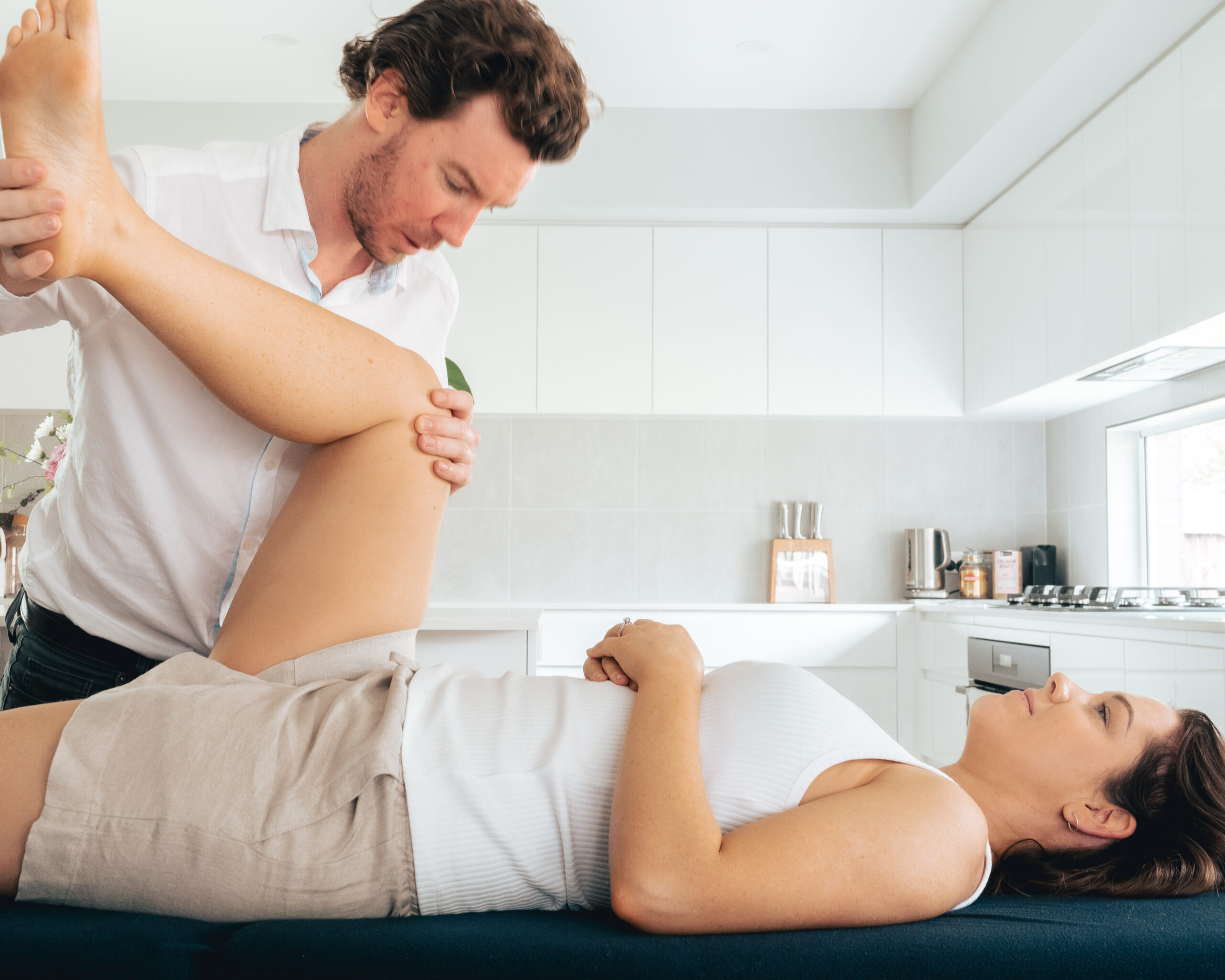
Physiotherapist assessing a client's hip movement on a treatment table
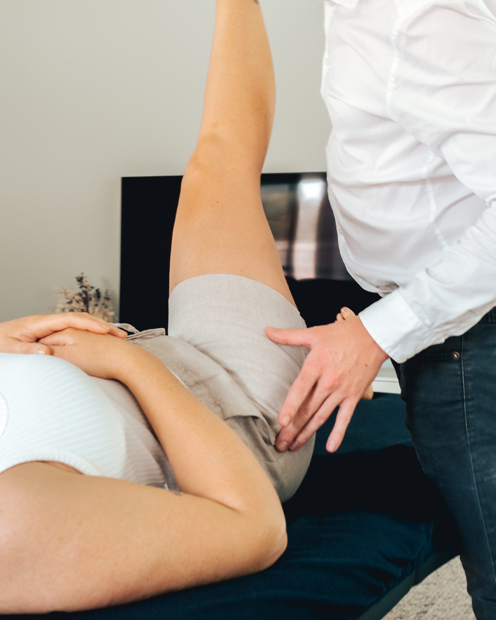
Physiotherapist assessing a client's hip with the leg raised

What is the anatomy of the hip?

The hip joint is a ball-and-socket joint, so the rounded head of the femur (thighbone) fits into the cup-shaped acetabulum of the pelvis. This type of joint allows for a wide range of motion, including flexion, extension, abduction, adduction, internal rotation, and external rotation.

The following bones form the hip joint:
Femur: The femur is the thighbone. The rounded head of the femur fits into the acetabulum of the pelvis.
Pelvis: The pelvis is a basin-shaped structure that forms the base of the trunk, and the acetabulum is a cup-shaped socket in the pelvis that receives the head of the femur.

The articulating surfaces of the hip joint are covered with hyaline cartilage, which helps to reduce friction and wear and tear on the joints. The joint capsule is a thin layer of tissue that surrounds the joint and helps to keep it in place. The femoral nerve also innervates the hip joint.
Diagram of the hip joint showing the ball and socket

Several ligaments, which are tough bands of tissue that connect the bones of the joint support the hip joint. The ligaments of the hip joint work together to stabilise the joint and prevent it from dislocating.

The three main ligaments of the hip joint are:

Iliofemoral ligament: This ligament is the strongest ligament in the body. It runs from the ilium (the uppermost bone of the pelvis) to the femur (the thighbone). The iliofemoral ligament prevents the hip from hyperextending (bending too far back).
Pubofemoral ligament: This ligament runs from the pubis (the bone that forms the front of the pelvis) to the femur. The pubofemoral ligament prevents the hip from abducting (moving away from the midline of the body).
Ischiofemoral ligament: This ligament runs from the ischium (the bone that forms the back of the pelvis) to the femur. The ischiofemoral ligament prevents the hip from rotating inward.

Besides these three main ligaments, there are also several smaller ligaments that help to stabilise the hip joint. These ligaments include the ligamentum teres (round ligament of the hip), the acetabular labrum, and the zona orbicularis.

The ligamentum teres is a small ligament that attaches the head of the femur to the acetabulum (the socket of the hip joint). The acetabular labrum is a ring of cartilage that surrounds the acetabulum. The zona orbicularis is a group of fibres that surround the joint capsule.

These ligaments work together to stabilise the hip joint and prevent it from dislocating. However, even with these ligaments, the hip joint is still a relatively unstable joint. This is because the head of the femur is much larger than the acetabulum. This allows for a wide range of motion in the hip joint, but it also makes the joint more susceptible to dislocation.
Diagram of the ligaments at the front of the hip joint

Several muscles, which work together to move the thigh and leg support the hip joint. We can divide the muscles of the hip joint into two groups:

Hip flexors: These muscles flex the hip joint, bringing the thigh towards the chest.
Hip extensors: These muscles extend the hip joint, straightening the thigh.
Hip abductors: These muscles abduct the hip joint, moving the thigh away from the midline of the body.
Hip adductors: These muscles adduct the hip joint, moving the thigh towards the midline of the body.
Hip internal rotators: These muscles rotate the thigh inward.
Hip external rotators: These muscles rotate the thigh outward.
Some of the most important muscles of the hip joint include:

Iliopsoas: This is the strongest hip flexor. It comprises two muscles, the psoas major and the iliacus. The iliopsoas originates from the lumbar spine and the iliac fossa of the pelvis. It inserts into the lesser trochanter of the femur.
Gluteus maximus: This is the largest muscle in the body. The gluteus maximus originates from the ilium, the sacrum, and the coccyx. It inserts into the iliotibial band and the greater trochanter of the femur.
Hamstring muscles: These muscles are on the back of the thigh. They comprise the semitendinosus, the semimembranosus, and the biceps femoris. The hamstring muscles originate from the ischial tuberosity. They insert into the tibia and the fibula.
Rectus femoris: This is one of the four quadriceps muscles of the thigh. It is on the front of the thigh. The rectus femoris originates from the ilium. It inserts into the patella and the tibial tuberosity.
Adductor magnus: This is the largest of the adductor muscles. It is on the medial side of the thigh. The adductor magnus originates from the pubis, the ischium, and the femur. It inserts into the linea aspera of the femur.

The muscles of the hip joint work together to allow us to walk, run, climb stairs, and perform other movements that require the movement of the thigh and leg. They also help to stabilize the hip joint and prevent it from dislocating.
Diagram of the hip and thigh muscles from the front and back

Hip Pain Subjective Assessment

Determining the history of your hip pain and how it all began and has evolved over time is crucially important. Without an understanding of how everything started and what set it all off, or in some cases what didn't happen, we won't quite know where to begin with objective testing.

What Happened?

As with every other pain presentation, I start with how your symptoms began. As I mentioned above, for some people this could have been months or even years ago, but for me when I saw someone it was maybe two days after it started, so I could really pinpoint how it started. If you can’t though, that’s totally cool because not everybody has an instigating incident. Usually in these cases I may replace my question with what is the first thing you remember about the pain, this can be a really important question because it’s often the most significant moment that the client can recall rather than just the first moment.

How does it behave?

Next, I want to understand how your symptoms have changed and developed over time and how the symptoms behave throughout the day. Changing symptoms over time, such as an increase in tightness or stiffness to a certain area, can help show what compensations might happen as the body’s strategy to get away from the pain. Other people may feel that their other hip gets sore because it’s compensating (hint: that’s when it’s really time to get on top of something and not “wait and see what happens”). How the pain behaves throughout the day is also highly valuable information. Whether activities such as stairs, walking up an incline, squatting down, sitting for prolonged periods of time or sleeping aggravate your symptoms can give us superb insights into your pain and what may cause it. It also gives us a wonderful insight into how we can structure an exercise program that both fits into your daily life and doesn’t aggravate your symptoms while allowing you to continue to live the life you want to and choose to. An exercise program shouldn’t become the be-all and end-all. You still need to enjoy life.

Any Strange Sensations?

Next up is determining whether you have any symptoms, such as clicking, catching, or locking sensations. This might indicate something happening within the hip joint, so these are important symptoms to pass on if you are experiencing them.

Injury History

The only other major factor left for us to discuss will be whether you have experienced any hip pain prior to this episode. A prior history of hip injuries or pain might indicate there is more going on than what it may look like on the surface.

Hip Pain Objective Assessment

The hip joint is a little like the Bermuda Triangle, very easy to get lost and not know where you are or what's going on. Simplicity is often the best approach. Utilising the Doha Agreement and its five clinical entities is the research recommended approach to objective assessment of hip and groin pain when coupled with a functional assessment.

A Testing Battery is Better than Single Tests

Assessing to differentially diagnose hip pain can be a tedious process. There are a lot of structures to rule out, ranging from the lower back to the hip joint, to ruling out muscular or tendon presentation, and nerve pain presentations. There’s a lot going on in such a small area. As I mentioned above, a few years ago, there was a consensus agreement on the definition and diagnostic criteria to assist clinicians in diagnosing hip and groin pain. The testing battery is short and concise, with two to three tests for each entity. Once this has been narrowed down or ruled out the entities, we can go a bit more in-depth with our testing.

Joints and Muscles

Specifically looking at the hip joint, we are often looking at testing to stress the capsule around the hip joint or the surface of the socket of the joint. There’s a range of tests that I can perform to apply different stresses. Positive tests are indicative that there could be something going on with the hip joint. They aren’t exactly the best tests at being specific. However, if you have positive tests coupled with painful clicking or clunking sensations, my clinical suspicion of it increases.

For muscular tests, we are often wanting to perform isometric assessments of the muscles to isolate the contraction of the muscle and have as few other contributing factors in the test. Often I will perform an assessment of the hip flexor and adductors as standard, but I may branch out with other tests should I feel the clinical need and depending upon the area of symptoms.

One of my other favourite hip tests to perform is the Modified Thomas Test. This is a great test because it allows us to assess the length of a range of unique structures ranging from the hip flexors, to the quads, to adductors, and the lateral hip complex, including the glutes and tensor fascia latae.

Greater Trochanteric Pain Syndrome

For a more specific diagnosis, such as a greater trochanteric pain syndrome, where the pain is quite localised to the side of the hip. There is again a specific testing process I choose to run through. The testing combines tests that are both specific and sensitive. What that means is that if I am turning up consistent positive results, my clinical suspicion rises significantly.

Functional Tests

Once we move beyond the clinical assessment procedure, we move on to the functional aspect of the assessment. This is really important, particularly for people who are having functional concerns with movements such as stairs, squatting, prolonged standing, or running. We will run through specific movement patterns to load and assess the capacity of the hip and pelvis to absorb the load adequately. From this, we can determine any areas of potential weakness or lack of motor control where we can develop an exercise program to address them.

Steps to Success for Rehabbing a Hip Injury

Treating hip pain shouldn’t be too difficult if you come to a concise and accurate diagnosis. However, sometimes where the diagnosis may be a bit of a battle between two different choices, I will treat one pathway and treat it well and then assess the response. If I were to treat both, if you get worse, I don’t know which treatment pathway made you worse. Conversely, if you get better, I also don’t know which one made you better. So I always err on the side of putting all my eggs in one basket and then reassessing afterwards, treating two or more areas in an initial consult has almost always resulted in myself and my client getting confused.

Hands On Treatment

Soft tissue release is probably my go-to treatment option around the hip joint. Releasing muscular tension can help to free up some movement around the hip and make movement throughout the day that bit easier. The most common areas that I find I am working on are the glutes, tensor fascia latae, and the hip flexor and quadriceps.

Another set of common techniques I will opt for is hip joint mobilisations, I find traction techniques of the hip joint can be incredibly effective for clients who are suffering from a hip joint-based presentation, whether it be osteoarthritis or a more acute injury such as a labral tear that a surgeon wants to trial non-surgical management on. Dry needling can also be highly effective around the hip joint in allowing us to get an isolated and specific effect on some of the deeper gluteal and hip rotator musculature.
Physiotherapist assessing the side of a client's hip

Hip Physio Exercises

As with everything else physiotherapists treat, a management plan isn’t complete without an individualised exercise program. For hips, these are so important and I really don’t think you can truly rehabilitate a hip injury without an exercise program, there will be too many movement adaptations and potential areas of weakness that I think your likelihood of another injury or insidious onset of pain will be too high. Most of the work you should do should be strengthening work around the gluteals, hip flexors, core, and abdominals. Developing the control and endurance of these muscles and power for specific movements and sports is vital for either returning to sport or just being able to maximise your quality of life.

Your Questions About Hip Pain

What causes hip pain?

Where your pain is gives the biggest clue. Pain on the outside of the hip is most often gluteal tendinopathy, sometimes with bursitis. Pain deep in the groin or front of the hip is more likely to come from the hip joint itself, such as osteoarthritis, a labral tear or hip impingement. Pain in the buttock can come from the lower back or the hip. Muscle strains around the hip and groin are common in sport. My assessment works through these possibilities so we know exactly what we are treating.

Is my hip pain arthritis?

Hip osteoarthritis usually causes pain in the groin or front of the hip, sometimes spreading to the thigh or knee. It is often accompanied by stiffness first thing in the morning or after sitting, and difficulty with tasks like putting on socks, getting in and out of the car or cutting your toenails. It is more common after 45 years of age or after a previous hip injury. Pain on the outside of the hip is less likely to be arthritis. A clinical assessment can usually tell, and an x-ray can help confirm it if needed.

Is it gluteal tendinopathy or bursitis?

Often it is both. Pain on the outside of the hip used to be called trochanteric bursitis, but research shows the main problem is usually the gluteal tendons, which attach to the bony point of the hip. The bursa sitting over them can become irritated as well. Both cause pain lying on your side, climbing stairs, standing on one leg and sitting with your legs crossed. The good news is they are managed in a similar way, with load management and a progressive strengthening program for the glutes.

What exercises help hip pain?

It depends on the cause, but strengthening the muscles around the hip is the foundation for most hip pain. For gluteal tendinopathy, that often starts with gentle holding exercises like pushing your knee out against a band, then progresses to bridges, side steps, step-ups and single-leg work. For hip osteoarthritis, strength and movement exercises plus walking or cycling help reduce pain and keep you active. Some common stretches, like crossing your leg over to stretch the outside of the hip, can aggravate tendon pain, so I tailor your program to your diagnosis.

Will I need a hip replacement?

Many people with hip osteoarthritis manage well for years without surgery. Exercise, education and weight management, where relevant, are the recommended first steps, and they can help reduce pain and improve function. A hip replacement is usually considered when pain is severe, sleep and daily life are significantly affected, and good non-surgical care has not helped enough. If you do head towards surgery, being as strong as possible beforehand helps your recovery, and I also provide physio after surgery at home.
Physiotherapist treating the front of a client's hip

Do you need a hip scan?

Honestly, I don’t find there are many cases of hip pain that need a scan. Sometimes they can come in handy, but more often than not, we can come to a relatively clear diagnosis and treatment pathway. Most times I may refer to a GP for imaging in the cases that I suspect there may be an issue within the joint, or I believe referral to a surgeon may be recommended, or other avenues such as a cortisone injection could be of benefit.

Realistically, the form of imaging that will be of benefit depends entirely on your symptoms and what we are trying to rule in or out. For issues within the joint, such as a labral tear, an MRI would be your best option. If we are assessing a case of greater trochanteric pain syndrome, ultrasound will be your best bet, an MRI doesn’t give significantly more information, and most times, we are looking at the bursa to assess whether a cortisone injection will be of benefit. An x-ray could be used in a few circumstances, bone stress injuries are a possibility though I may lean towards getting a CT scan for those, often I would refer for an x-ray in more of a paediatrics population to assess for any potential femoral head concerns.

Returning to Training and Sport Post Hip Injury

Getting back on the sporting field is every athlete’s goal. I strive to send each athlete back on the sporting field a more complete and robust athlete than what they were when they got injured. Returning to training and inevitably getting back to playing competitive games is a deliberate and planned process that needs to be mapped out from start to finish. Too often I hear of clients developing their own return to sport plan that often goes from “Yeah I’ll rest up” straight to “Yeah I’ll play a full game this week coach”. It’s better than I’ll just play through the pain, but it can be a lot better than that.

Returning to training needs to be a graded progression. Often in the first session, I will get my clients to do some light running and agility work, take part in the warm-up and complete some sport-specific movements or skill-based work. Assuming they respond positively to that, we can broaden our horizons from there. Often I will work to increase the speed and individual difficulty of drills an athlete is performing before I then return them to gameplay scenarios and eventually full gameplay.

To return to sport, I have one major rule for athletes that I manage. They have to have completed at minimum one full training session prior to going back to play. Ideally, I would like to have had one full week of training, but sometimes, this may not always be reasonable. For example, a client undergoing a two-week return to sport plan will have four progressions, assuming they train twice per week. It’s rare that I will progress to full training by the third session, so the fourth will become the only full training session we have planned. Whereas an athlete undergoing a three-week plan, most times we will manage it so that the third week is a full training week.
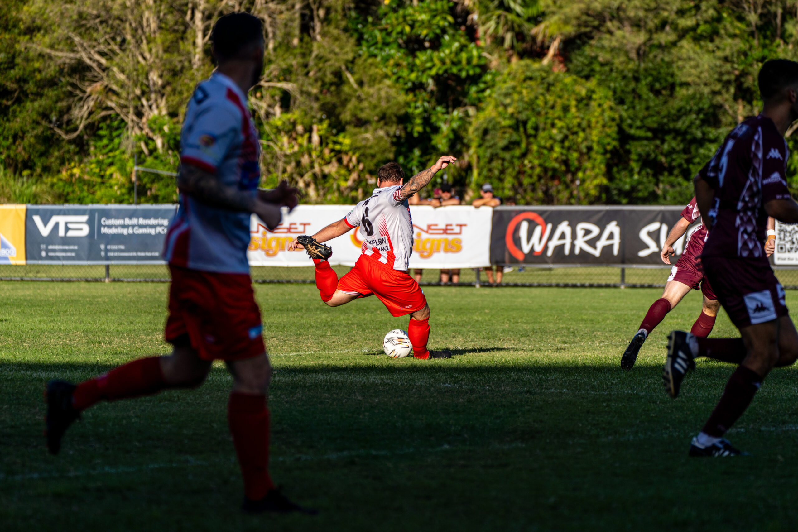
Footballer taking a shot during a local match

Hip Pain FAQs

Muscle strains and irritated tendons around the hip commonly improve within 4 to 8 weeks with the right exercise program. Longer-standing problems such as gluteal tendinopathy or pain from within the hip joint may take 3 months or more. I will give you a realistic estimate after your assessment.

Pain when lying on your side at night is common with gluteal tendinopathy and trochanteric bursitis, where the weight of your body compresses the sore tendons on the outside of the hip. A pillow between your knees, or lying on your back or other side, can help while we work on the cause.

Often yes, with some changes. Reducing your distance, avoiding hills and cambered surfaces, and shortening your stride can let you keep running while your hip settles. If running makes your pain worse the next day, we pull back and build up again gradually.

See your GP promptly if your hip pain started after a fall and you cannot put weight through the leg, if you have a fever or feel unwell, or if the pain is severe at night and getting steadily worse. In children and teenagers, a new limp or hip pain should always be checked.

Each consultation is 45 minutes and costs $110, for both your first appointment and follow-ups. Dry needling, taping and exercise bands are included at no extra cost. If you need a longer course of rehab, my 3, 6 and 12 week rehabilitation programs work out cheaper than paying per session.

Yes. You can claim a rebate from your health fund for every session. PRP Physio is not a preferred provider for any fund, so your rebate may be slightly smaller than at a preferred provider. Your fund can confirm the exact amount using item code 500 for your first consultation and 505 for follow-ups.

Yes, if your hip pain was caused by an injury or strain at work. Your GP starts the claim and gives you a WorkCover certificate, which acts as your referral. Once your claim is accepted, I bill WorkCover directly, so there are no out-of-pocket costs for you. WorkCover covers your first five sessions, and I speak with your case manager if you need more.

Mobile Physio: Get in Touch

I'm ready to book online now

For those who are ready to book an appointment right now, let's get started on your journey back to sport!

Just hit the button below and it will take you straight to an online booking platform. You will just have to enter a few details and it will all be booked in for you!

I'd rather have a conversation first

Some injuries are complex, should you see a GP first, should you go get a scan, is a physio even the right person to see? Sometimes a 10 minute conversation can be enough to answer some of these questions.

Just submit a contact form on the link below and I'll give you a call ASAP and we can chat through a few things to get you moving in the right direction!
© 2026 PRP Physio. All rights reserved.